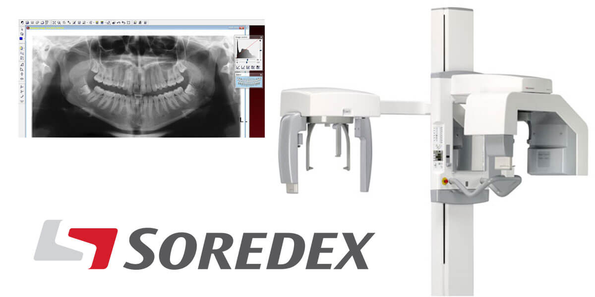
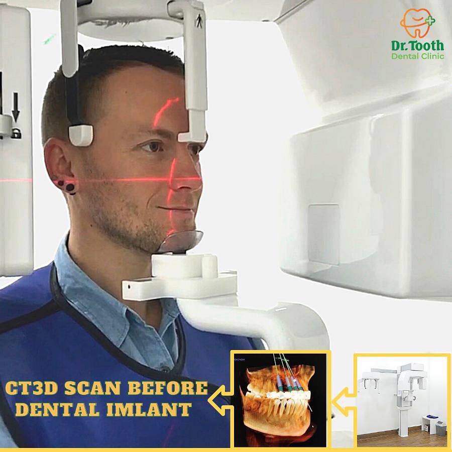
Improve diagnosis and treatment planning with comprehensive CBCT imaging platforms by Soredex. The doctor offers workflow efficiency and unsurpassed imaging quality, all while putting your patients first. Enjoy our dental 3D CBCT X-Ray machines for high quality images and highly comfortable patient positioning. Our CBCT machines offer 2D and 3D X-Ray dental imaging!
What is Panoramic X-ray?
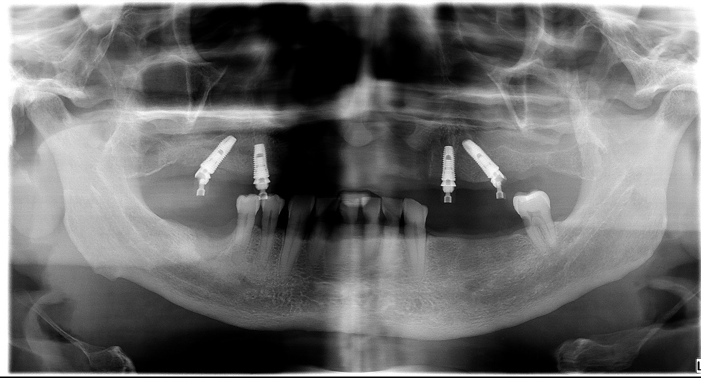
Panoramic radiography, also called panoramic x-ray, is a two-dimensional (2-D) dental x-ray examination that captures the entire mouth in a single image, including the teeth, upper and lower jaws, surrounding structures and tissues. The jaw is a curved structure similar to that of a horseshoe. However, the panoramic x-ray produces a flat image of the curved structure. It
usually provides details of the bones and teeth.
Panoramic dental x-ray uses a very small dose of ionizing radiation to capture the entire mouth in one image. It is commonly performed by dentists and oral surgeons in everyday practice and may be used to plan treatment for dentures, braces, extractions and implants.
A panoramic x-ray can also reveal dental and medical problems such as:
- Advanced periodontal disease
- Cysts in the jaw bones
- Jaw tumors and oral cancer
- Impacted teeth including wisdom teeth
- Jaw disorders
- Sinusitis
What is Dental Cone Beam CT?
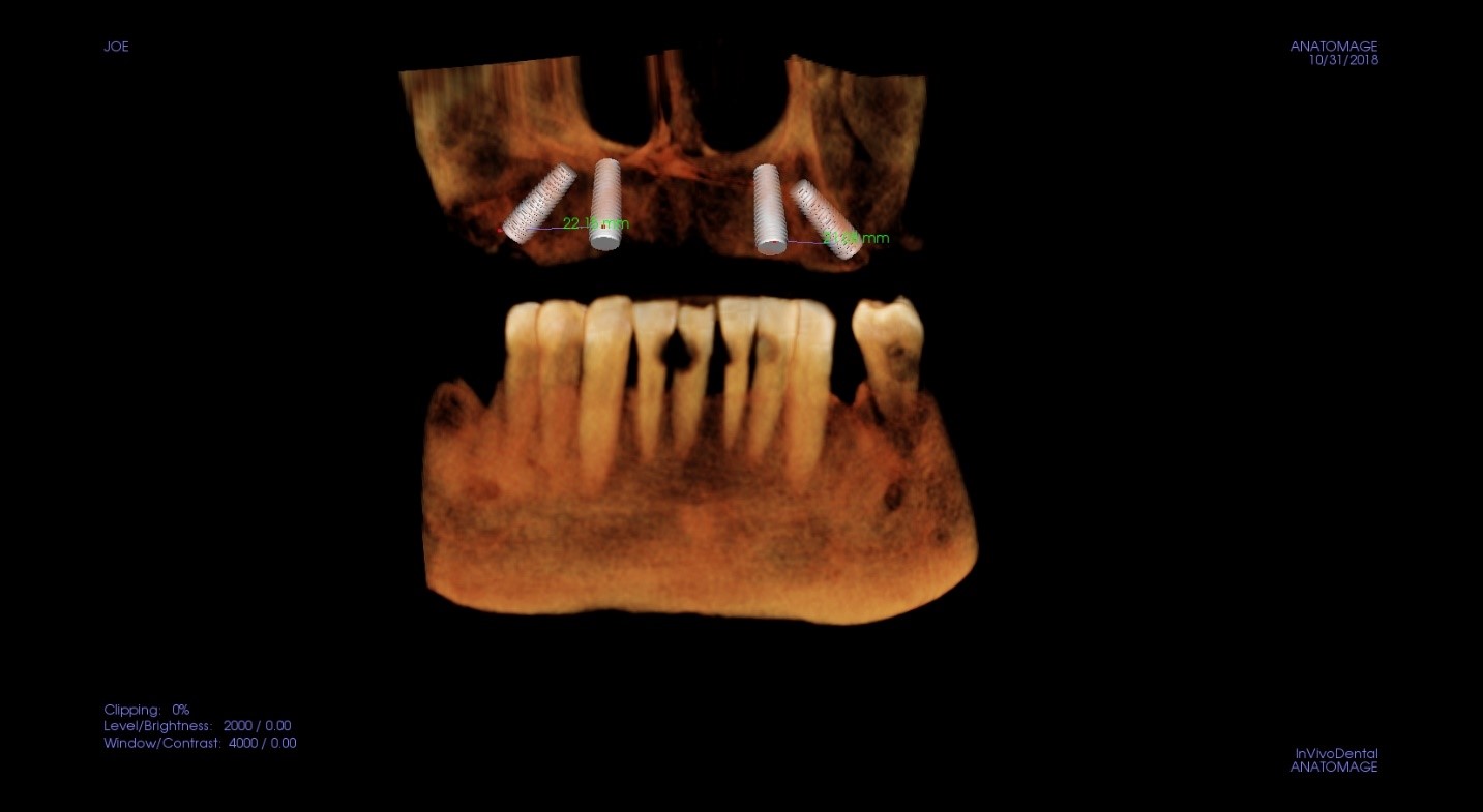
Dental cone beam computed tomography (CT) is a type of CT scanner uses a special type of technology to generate three dimensional (3-D) images of dental structures, soft tissues, nerve paths and bone in the craniofacial region in a single scan. Images obtained with cone beam CT allow for more precise treatment planning.
Cone beam CT is not the same as conventional CT. With cone beam CT, an x-ray beam in the shape of a cone is moved around the patient to produce a large number of images, also called views. CT scans and cone beam CT both produce high-quality images. It provides detailed images of the bone and is performed to evaluate diseases of the jaw, dentition, bony structures of the face, nasal cavity and sinuses. It does not provide the full diagnostic information available with conventional CT, particularly in evaluation of soft tissue structures such as muscles, lymph nodes, glands and nerves.
Dental cone beam CT is commonly used for treatment planning of orthodontic issues. It is also useful for more complex cases that involve:
surgical planning for impacted teeth.
diagnosing temporomandibular joint disorder.
accurate placement of dental implants.
determining bone structure and tooth orientation.
locating the origin of pain or pathology.
cephalometric analysis.
REMARK: You should wear comfortable, loose-fitting clothing to your exam. You may be
given a gown to wear during the procedure.
Metal objects, including jewelry, eyeglasses, dentures and hairpins, may affect the CT images and should be left at home or removed prior to your exam. You may also be asked to remove hearing aids and removable dental work. You may be asked to remove any piercings.
You can review our clinics here:
DR. TOOTH DENTAL CENTER – 90 Huynh Thuc Khang, Nha Trang
Thank you for choosing us!
Hotline: 0964 888 679 - 0813 515 788
Opening time: 8h00 – 18h00 (Mon- Sat).
Fanpage: Nha khoa DrTooth
Make appoitment: Đặt lịch hẹn tư vấn miến phí